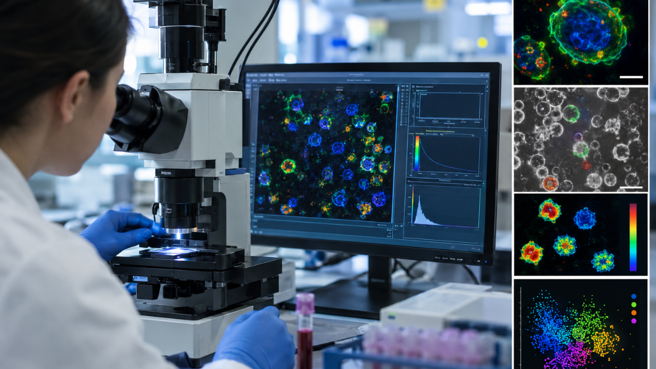
Traditional laboratory science has relied on chemical stains and fluorescent dyes for decades to figure out what our blood cells are doing. It is messy, expensive, and alters the very biology you are trying to measure. Now, researchers have published a study in Biophotonics Discovery outlining a method to bypass chemical dyes entirely. By reading natural cellular light, scientists can spot activated immune cells from ordinary blood samples with nearly 94 percent accuracy in just two hours.
If you work with cellular diagnostics or cell-based therapies, this changes the operational playbook. Let's break down how this works and why it matters.
The Problem With Traditional Dyes
For years, spotting active immune cells meant coating them in fluorescent antibodies or chemical markers. These labels require heavy sample preparation, extra reagents, and plenty of hands-on laboratory time. Worse, adding foreign compounds can alter cell behavior or kill the sample before you finish the analysis.
You end up looking at a static snapshot. You know what cell type is present, but you miss the live functional state. Biology does not pause just because you added a dye.
Reading Natural Cellular Glow
Instead of sticking artificial markers onto cells, the new approach looks inward. It relies on optical metabolic imaging (OMI) using two-photon microscopy.
Cells naturally produce light through internal molecules involved in cellular metabolism. This phenomenon is known as autofluorescence. By measuring how long those internal molecules stay in an excited state before returning to normal—known as fluorescence lifetime—researchers map out active metabolic shifts.
In tests using peripheral blood mononuclear cells (PBMCs) from three healthy donors, thousands of individual cells were scanned under resting and stimulated conditions. Machine learning models then parsed the data. The results surprised even the researchers. The imaging technique distinguished activated cells from resting cells with nearly 94 percent accuracy just two hours after stimulation.
Where Individual Cell Populations Stand
Not all immune cells behave identically under an optical scanner. The study revealed distinct metabolic signatures across different populations:
- Monocytes: Part of the innate immune system, these rapid responders were identified with 96 percent accuracy in resting samples and 88 percent accuracy after activation.
- Natural Killer (NK) Cells: These showed about 74 percent accuracy across both resting and activated conditions.
- T Cells and B Cells: These adaptive immune populations displayed more similar metabolic profiles, showing that standard identity markers still offer valuable context when combined with metabolic data.
This metabolic lens proves that two cells looking identical under a standard microscope can have completely different functional states.
Practical Applications for Modern Labs
What does this mean for day-to-day research? Non-destructive imaging transforms how labs assess cell fitness prior to advanced processing. In cell-based therapies, knowing the exact metabolic health of individual cells without contaminating them with chemical reagents reduces pipeline failure rates.
Stop relying solely on surface stains when analyzing functional changes. Incorporate label-free optical methods early in your experimental designs to preserve your samples and capture real-time metabolic shifts.